
SIS biomaterials with independent intellectual property rights, the main components include Collagen I, III, fibronectin and a variety of growth factors (TGF-β, VEGF and FGF, etc.). Growth factors are cell factor secreted by a variety of cells, acting on specific target cells, regulating cell division, matrix synthesis and tissue differentiation, and contributing to the enhancement of cell function and the formation of vascularization.
This paper focuses on the quantitative detection of three growth factors (TGF-β, VEGF and FGF) in SIS materials, and the detection results are obtained by two experimental methods. The article was published in the journal Materials Science in May 2018. The following are excerpts.

Figure: Cover of the Journal Materials Science in May 2018
Introduction to growth factors
TGF-β, a rich bone matrix protein, is also a peptide anti-inflammatory growth factor. It is widely present in normal tissues and transformed cells, and has a high content in platelets and bone tissue. It plays an extremely important role in bone formation and bone remodeling. Immune regulation can be carried out by controlling the proliferation, differentiation and activation of immune cells (T cells, B cells, etc.) to promote wound healing and vascular growth.
VEGF, a vascular-derived growth factor with high specificity to vascular endothelial cells, is synthesized and secreted by platelets, vascular smooth muscle cells, endothelial cells, osteoblasts or tumor cells, etc. It is essential for the formation of new blood vessels, has functions such as increasing vascular permeability and promoting endothelial cell proliferation, and also has the effect of protecting nerves.
FGF, a group of polypeptides with similar structural characteristics, is widely present in body tissues, has a strong role in promoting cell division and proliferation, and can induce the formation of capillaries.

Figure: Animation diagram. SIS material is rich in collagen I, III, fibronectin and various growth factors (TGF-β, VEGF and FGF, etc.)
Introduction to extraction method
Method 1: Weigh 5mg of the crushed SIS material, add 1mL of Buffer 1, homogenize at high speed and low temperature for 20-30min until the liquid becomes a colloid, centrifuge at 14,000rpm for 30min, extract the supernatant, dilute to 1mL, and store at 4℃.
Method 2: Weigh 5mg of the crushed SIS material, add 1mL of Buffer1, homogenize at high speed and low temperature for 20-30min until the liquid becomes colloidal, stir at a constant speed at 4℃ for 24h, centrifuge at 14,000rpm for 30min, extract the supernatant, dilute to 1 mL, and store at 4℃.
ELISA detection method
Double Antibody Sandwich ELISA (DAS-ELISA) was used for TGFβ1 and VEGF, and Competitive Inhibition ELISA was used for FGF2.
The samples treated by the two methods were operated according to the instructions of the ELISA kit, and the operation steps were as follows (taking TGFβ1 as an example):
1) Add sample: set Standard well, Sample well, Blank well respectively, add 100μL of standards and samples of different concentrations in turn, add standard buffer to Blank well. Cover plate and incubate at 37℃ for 1h after. Remove the liquid and spin dry;
2) Add 100μL of detection solution A working solution to each well, incubate at 37℃ for 1h, remove the liquid, wash each well with 350μL of washing solution for 1-2 min and spin dry, this process is repeated 3 times;
3) Add 100μL of detection solution B working solution to each well, incubate at 37℃ for 1h, remove the liquid, wash each well with 350μL of washing solution for 1-2 min and spin dry, this process is repeated 5 times;
4) Add 90μL of TMB Substrate Solution to each well, develop color at 37℃ avoid direct exposure to intense light (10-20min), add 50μL of Stop Solution to each well to stop the reaction, and shake gently;
5) Immediately use Microplate Reader to measure the optical density value (O.D. value) of each well at a wavelength of 450nm;
6) Establish a standard curve according to the standard product, substitute it into the formula to obtain the content of active growth factor in the sample, and then convert it into the content in the dry weight of the SIS graft sample.
Test results
TGFβ1 test results
According to the optical density value (O.D. value) of TGFβ1 standard substance of different concentration in ELISA kit at 450nm wavelength, a standard curve is made, as shown in Figure 1.

Figure 1: Standard curve of O.D. values of different concentrations of TGFβ1 standard
As shown in Figure 2, the content of TGFβ1 in SIS detected by method 1 was 5391±370 pg/g, and the content of TGFβ1 in SIS detected by method 2 was 8375±2125 pg/g.

Figure 2: Comparison of TGFβ1 assay results between the two methods
VEGF test results
According to the optical density value (O.D. value) of VEGF standard of different concentrations of the ELISA kit at a wavelength of 450nm, a standard curve was made, as shown in Figure 3.

Figure 3: Standard curve of O.D. values for different concentrations of VEGF standards
As shown in Figure 4, the content of VEGF in SIS detected by method 1 was 3974±871 pg/g, and the content of VEGF in SIS detected by method 2 was 5486±1043 pg/g.

Figure 4: Comparison of VEGF detection results between two methods
FGF2 test results
According to the optical density value (O.D. value) of FGF2 standard of different concentrations of the ELISA kit at a wavelength of 450nm, a standard curve was made, as shown in Figure 5.


Figure 5: Standard curve of O.D. values for different concentrations of FGF2 standards
As shown in Figure 6, the content of FGF2 in SIS detected by method 1 was 3990±1372 pg/g, and the content of FGF2 in SIS detected by method 2 was 2668±384 pg/g.

Figure 6: Comparison of FGF2 detection results between two methods
Conclusion
Growth factors have very strict requirements on extraction conditions. Different growth factors have different suitable extraction methods due to their different types and mechanisms of action. In this experiment, by comparing the pretreatment methods of growth factors of SIS materials, the most suitable extraction methods for three growth factors (TGFβ1, VEGF, FGF2) were obtained and their contents were detected. Among them, the content of TGFβ1 in Biosis Healing SIS was 8375±2125 pg/g, the content of VEGF was 5486±1043 pg/g, and the content of FGF2 was 3990±1372 pg/g.
Wound healing is a complex biological process, and growth factors play an important role in improving various physiological and pathological states, promoting tissue regeneration, repair and wound healing processes. While promoting the formation of granulation tissue, it accelerates the process of wound re-epithelialization.
The SIS material with independent intellectual property rights of Biosis Healing has the advantages of good biocompatibility, promotion of tissue regeneration and repair, accelerated angiogenesis, and resistance to infection. It is widely used in the repair and reconstruction of tendons, dura mater, abdominal wall, skin, oral cavity and other tissues.
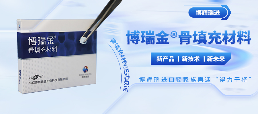
博瑞金® Oral Biological Graft, the first SIS material oral repair membrane in China, is a powerful guarantee for perfect soft tissue repair and efficient osteogenesis. The product is designed with a patented technical structure. After being implanted into the damaged area of the oral cavity, it can effectively play a protective role as a barrier, realize the rapid ingrowth of blood vessels, and induce tissue repair and regeneration.
Compared with inert tissue-derived materials such as dermis on the market, 博瑞金® Oral Biological Graft has good softness, fit and hydrophilicity, which can better fit the defect area and facilitate surgical operations; is rich in bioactive substances which can promote cell attachment and tissue regeneration; has excellent tolerance to infection, can reduce the chance of wound infection.

Figure: SIS material 博瑞金® Oral Biological Graft
博瑞金® Oral Biological Graft has been on the market, and the national investment promotion has been officially launched. For more product details, please contact us.
Fax:010-61252660-892


版权所有®2019BIOSISHEALING


